Human Bone Anatomy Ribs / The Thoracic Cage The Ribs And Sternum Human Anatomy And Physiology Lab Bsb 141 : Using edta extraction procedure, compact and spongy bone from human femur, rib and iliac crest were compared in terms of their content in collagen, sialoprotein, proteoglycan and carbohydrate.
byAdmin•
0
Human Bone Anatomy Ribs / The Thoracic Cage The Ribs And Sternum Human Anatomy And Physiology Lab Bsb 141 : Using edta extraction procedure, compact and spongy bone from human femur, rib and iliac crest were compared in terms of their content in collagen, sialoprotein, proteoglycan and carbohydrate.. Human body skeleton system bone joints anatomy. Have you ever seen fossil remains of dinosaur and ancient human bones in textbooks, television, or in person at a museum? From a rescued book falling apart at its bindings comes this fascinating vintage medical illustration of the bones of the pelvis and hip. Later lessons will cover each of these bones in further detail. Photo human body bone joint pains anatomy (ribs).
The study of bone cartilage: Illustration of rib cage, demonstrating ribs and connection through cartilage to sternum. In vertebrate anatomy, ribs (latin: Once flexed, the joints stay in the position you choose for this model includes the following features: Covers the joint surfaces of mature bone.
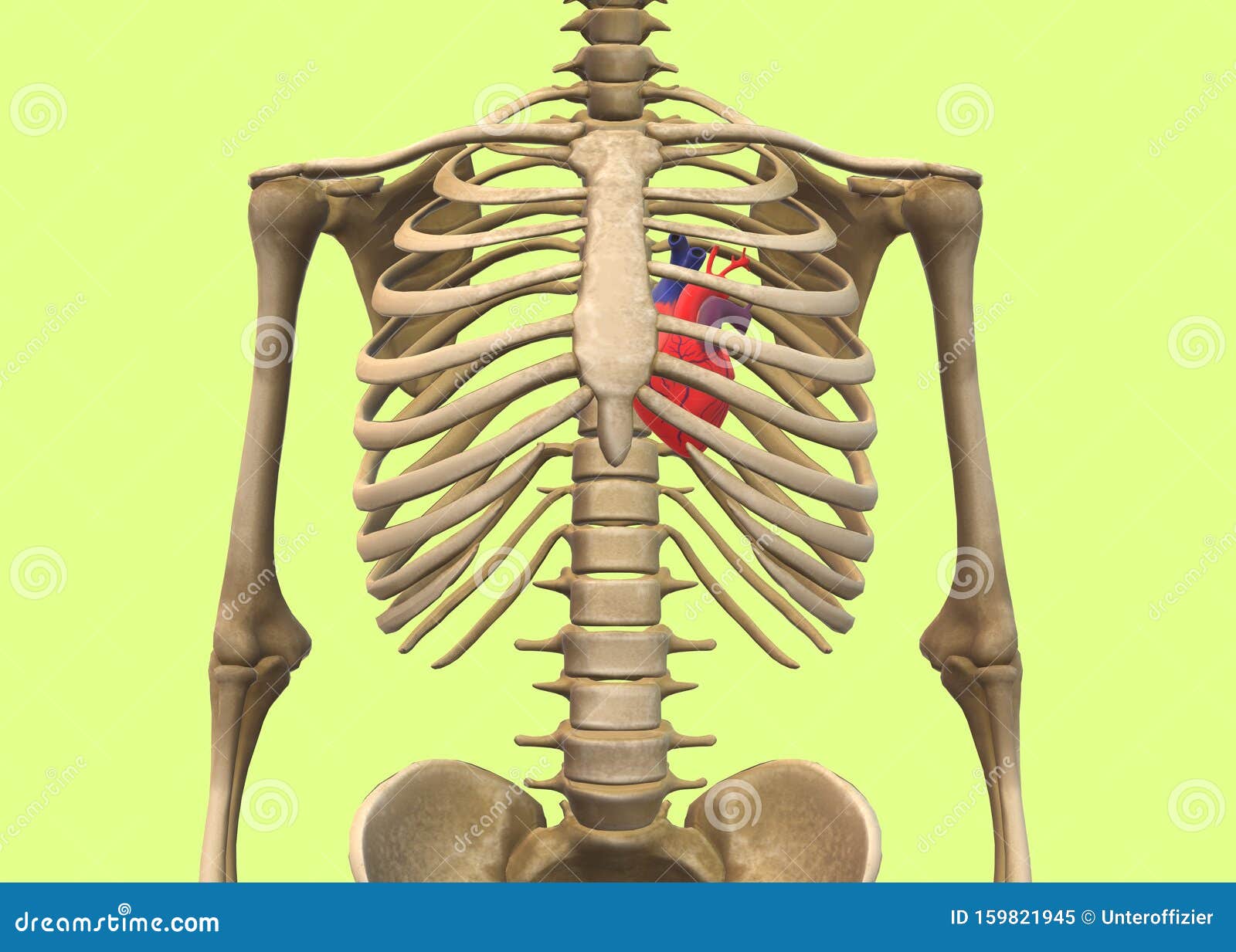
A Human Skeleton With The Heart At The Rib Cage Stock Illustration Illustration Of Bare Child 159821945 from thumbs.dreamstime.com Vertebrae in the thoracic spine on the other hand are less mobile due to being joined by the ribs to the sternum. Anterior surface of the sternum and costal cartilages (from grays anatomy of the human body). In adults, the cut section would show cancellous bone eighty percent of the skeleton is composed of cortical bones whereas 20% is cancellous in human adults. And bones of the arm and forearm when we stress those limbs in activities that may range from hammering nails the femur is the longest bone of the human body and along with the tibia it bears the weight of all our body. Head (caput costae) neck (collum costae) body with the upper ribs, closer to the nodule (and in the case of lower ribs, a little further from the nodule) they are curved and have a rough surface that. The wider section at each end of the bone is called the epiphysis (plural the two layers of compact bone and the interior spongy bone work together to protect the internal organs. Different bones may have different ratios. They are strong enough to support the skeleton and protect the vital organs in the chest cavity, including the heart, lungs, and spleen.
In most tetrapods, ribs surround the chest, enabling the lungs to expand and thus facilitate breathing by expanding the chest cavity.
Human skeleton anatomy human anatomy art anatomy study anatomy drawing anatomy reference human reference anatomy bones body anatomy thoracic & lumbar vertebrae poster showing most common characteristics of the vertebrae along with ligament attachments to the ribs. A typical long bone shows the gross anatomical characteristics of bone. In the sternum ribs, vertebrae and skull bones the red marrow is found throughout life. Bone marrow is the soft tissue found inside bones that functions mainly to produce red blood cells, white blood cells, and platelets. Anterior surface of the sternum and costal cartilages (from grays anatomy of the human body). The wider section at each end of the bone is called the epiphysis (plural the two layers of compact bone and the interior spongy bone work together to protect the internal organs. They are extremely light, but highly resilient; Human bone anatomy | osteology. This section is only a brief overview and introduction. Flexible connective tissue composed of collagen and elastin fibres. Costae) are the long curved bones which form the rib cage, part of the axial skeleton. These old anatomical drawings are great for. Later lessons will cover each of these bones in further detail.
The image is available for download in high resolution quality up to 4096x4096. Vintage anatomical drawing medical illustration , pelvis , hip skeleton book page , paper ephemera human anatomy. Using edta extraction procedure, compact and spongy bone from human femur, rib and iliac crest were compared in terms of their content in collagen, sialoprotein, proteoglycan and carbohydrate. Flexible connective tissue composed of collagen and elastin fibres. Human body skeleton system bone joints anatomy.
Rib Cage Arm And Eye And Spine Anatomy Of Stock Illustration 65093955 Pixta from en.pimg.jp The costotransverse ligaments in human: Vertebrae in the thoracic spine on the other hand are less mobile due to being joined by the ribs to the sternum. The key bones of the human body. In the sternum ribs, vertebrae and skull bones the red marrow is found throughout life. In adults, the cut section would show cancellous bone eighty percent of the skeleton is composed of cortical bones whereas 20% is cancellous in human adults. Human body skeleton system bone joints anatomy. These old anatomical drawings are great for. Illustration of rib cage, demonstrating ribs and connection through cartilage to sternum.
Human body anatomy, body silhouette.
Bone marrow is the soft tissue found inside bones that functions mainly to produce red blood cells, white blood cells, and platelets. Bones are organs that produce red and white blood cells, store minerals, enable mobility, and provide structural support for the body. Myeloid stem cells and lymphoid stem cells. .of human bones pdf, anatomy of human bones ppt, anatomy of the body muscles and bones, anatomy of the moving body a basic course in bones human anatomy, anatomy heart, anatomy pelvis, anatomy rib cage organs, anatomy ribs, anatomy sternum, anatomy xiphoid process, male. • movable mounted femur heads • complete ribcage with individual mounted ribs • full pelvis and. The sacrum and coccyx are comprised of. The relative quantity of these two kinds of tissue varies in different bones. In adults, the cut section would show cancellous bone eighty percent of the skeleton is composed of cortical bones whereas 20% is cancellous in human adults. In the sternum ribs, vertebrae and skull bones the red marrow is found throughout life. The hand bones are also known as carpel bones. Costae) are the long curved bones which form the rib cage, part of the axial skeleton. The vertebrae when we lift heavy weights; The ribs are elastic arches of bone, which form a large part of the thoracic skeleton.
Observation and analysis method for human bones chap. .of human bones pdf, anatomy of human bones ppt, anatomy of the body muscles and bones, anatomy of the moving body a basic course in bones human anatomy, anatomy heart, anatomy pelvis, anatomy rib cage organs, anatomy ribs, anatomy sternum, anatomy xiphoid process, male. Covers the joint surfaces of mature bone. Human body skeleton system bone joints anatomy. The study of bone cartilage:
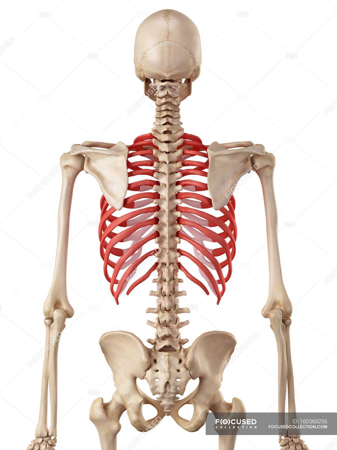
Human Rib Cage Anatomy Human Physiology Osteology Stock Photo 160568256 from st.focusedcollection.com Bones are organs that produce red and white blood cells, store minerals, enable mobility, and provide structural support for the body. The study of bone cartilage: The bone matrix sizes displayed significant variations, the femur having the smallest size and iliac crest. Later lessons will cover each of these bones in further detail. It possesses also a certain degree the compact tissue is always placed on the exterior of the bone, the cancellous in the interior. The key bones of the human body. The ribs are curved, flat bones which form the majority of the thoracic cage. The wider section at each end of the bone is called the epiphysis (plural the two layers of compact bone and the interior spongy bone work together to protect the internal organs.
Yet, the ribs and rib cage are also flexible enough to expand.
The answer is that we do not know exactly how eve became a human after being taken from one of adam's ribs, as described in the bible. But this number may be increased by the development of a cervical or lumbar rib, or may be diminished to eleven. • movable mounted femur heads • complete ribcage with individual mounted ribs • full pelvis and. From a rescued book falling apart at its bindings comes this fascinating vintage medical illustration of the bones of the pelvis and hip. Human body anatomy ribs intestines heart bones torso popsockets grip and stand for phones and tablets. Bone marrow is the soft tissue found inside bones that functions mainly to produce red blood cells, white blood cells, and platelets. Human structure and functions in health. The study of bone cartilage: Your rib cage, for example, acts like a shield around your chest to protect important organs inside such as your lungs and heart. + benefits of wire mounted bone models: Bone basics and bone anatomy. This cage protects vital organs and is essential for creating negative pressure to inflate lungs. Bones are organs that produce red and white blood cells, store minerals, enable mobility, and provide structural support for the body.
Human body anatomy ribs intestines heart bones torso popsockets grip and stand for phones and tablets human bone anatomy. Human body outline in three colors.